EMBRYOS

Preimplantation genetic testing proposed to predict risk of common diseases

Preimplantation genetic testing proposed to predict risk of common diseases

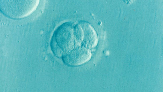
A new study claims to have mapped the genomes of embryos created by IVF and suggests that the same techniques used could also be used to enable preimplantation genetic testing (PGT).

The paper, published in Nature Medicine, outlines molecular and statistical techniques for inferring the whole genome sequence of three- and five-day-old in vitro embryos using tiny amounts of genetic material.

The authors believe that this makes it possible to forecast the risk of developing common diseases, such as cancer and diabetes, that are affected by many different genes. Such an approach to understanding the combined effects of many genes is called a polygenic risk score (PRS).

‘Our approach enabled the prediction of both rare and common variants in embryo genomes’ wrote the authors, from genomics company MyOme in California. ‘Our findings may inform the discussion of utility and implementation of genome-based PGT in clinical practice.’

To construct the genomes of more than 100 embryos, the researchers analysed hundreds of thousands of specific sites across the genome, in a process called genotyping. They filled in the gaps in the genomes with genetic sequences from the prospective parents, and compared their predicted genome to that of the born child.

The authors found that they were able, with 97-99 percent accuracy, to infer the correct sequence at sites used to calculate PRSs for 12 medical conditions. They say that this could enable the creation of PRSs for embryos.

While the method for assembling the genomes of embryos is of interest to the scientific community, the predictive value and ethical implications of so-called ‘PGT-P‘ (PGT that involves PGSs) are controversial.

‘It is important to stress that this study does not shed light on the most important aspect of using PRSs for embryo selection from IVF, where there are fundamental questions of both the statistical validity of the PRS in this setting and also the ethical appropriateness of procedure’, said Professor Ewan Birney, deputy director of the European Molecular Biology Laboratory, who was not involved in the study.

In the UK, selecting an embryo based on the genes it carries is only permitted for couples who are at risk of having a child with a serious genetic condition, such as Huntington’s disease or cystic fibrosis. The Human Fertilisation and Embryology Authority has recently stated that it would not be legal to use PGT-P for embryo selection in the UK.


https://www.nature.com/articles/s41591-022-01743-0


Placenta is the first organ to form in a fertilised egg

Placenta is the first organ to form in a fertilised egg

Placenta development is initiated first in human pregnancies, even before the embryo starts to form, according to new research.

A team of scientists at the Francis Crick Institute, London, has found that one of the first steps after egg fertilisation in mammals is the initiation of placenta creation, the organ responsible for providing oxygen and nutrients to the growing baby.

‘This study highlights the critical importance of the placenta for healthy human development,’ said Dr Kathy Niakan, senior author of the study and group leader at the Crick. ‘If the molecular mechanism we discovered for this first cell decision in humans is not appropriately established, this will have significant negative consequences for the development of the embryo and its ability to successfully implant in the womb.’

Once an egg is fertilised the cell rapidly divides and a key process called cell specialisation occurs, where each cell is assigned a specific task.

The team sought to examine the very first cell specialisations by studying donated human embryos that were surplus to in vitro fertilisation (IVF) treatment.

In embryos at the 16-32 cell stage, the team observed a subset of cells which changed shape and polarised, this triggered molecular events that drives placenta specialisation. In particular, atypical protein kinase C (aPKC) was highly expressed at one end of the cell, when aPKC was inhibited the cells no longer became placenta precursors.

This first cell specification is ‘widespread in mammals’ said researchers after they found the same result in cow and mouse embryos, which have divergent mechanisms at later stages of development.

This research, published in Nature, revealed that placenta development starts much earlier than previously thought, before the embryo has implanted into the wall of the uterus, suggesting placenta cells may have important functions in healthy embryo implantation and development.

During IVF treatment, a reliable predictor of successful implantation of an embryo is the presence of placental precursor cells under the microscope. Therefore, this work paves the way towards a better understanding of how to help those struggling to conceive.

‘Understanding the process of early human development in the womb could provide us with insights that may lead to improvements in IVF success rates in the future’ adds Dr Niakan, ‘It could also allow us to understand early placental dysfunctions that can pose a risk to human health later in pregnancy.’

Read more: https://www.nature.com/articles/s41586-020-2759-x

Source: https://www.bionews.org.uk/page_152173

Embryo vitrification is safe but longer storage reduces chances of pregnancy success

Embryo vitrification is safe but longer storage reduces chances of pregnancy success

Freezing and storing embryos during fertility treatment using a technique called vitrification is safe, although women are less likely to become pregnant and have a live birth the longer the embryos are stored, according to new research published in Human Reproduction, one of the world’s leading reproductive medicine journals.

There have been concerns that vitrification technology could be unsafe for the embryo, leading to complications at the time of birth and later, including preterm birth, low or high birthweight and birth defects.

Until now, it was also unclear whether storage time after vitrification affected embryo viability, pregnancy outcomes or neonatal outcomes. The study, which is the largest to investigate these questions, found that the chances of becoming pregnant and giving birth to a live baby significantly decreased with longer storage time.

The researchers from the Shanghai Ninth People’s Hospital (Shanghai, China) analysed outcomes from 24,698 patients who had vitrified embryos transferred for the first time between January 2011 and December 2017.

They compared patients who had vitrified embryos stored for up to three months (group one) with patients whose embryos were stored for 3-6 months (group two), 6-12 months (group three) and 12-24 months (group four). They found that the implantation rate fell from 40% in group one to 26% in group four; the clinical pregnancy rate fell from 56% in group one to 26% in group four; and the live birth rate fell from 47% in group one to 26% in group four.

This means that among women who had embryos stored for less than three months, 47 out every 100 women would achieve a live birth. Whereas among women who had embryos stored for between 12-24 months, 34 out of every 100 women would achieve a live birth.

The rate of miscarriages and ectopic pregnancies also increased with longer storage time. However, these associations were not statistically significant after taking into account factors that could affect the results such as the mother’s age, mother’s body mass index, the cause of infertility, parity and embryo quality and stage of development. There was no evidence that storage time affected neonatal outcomes.

Groups three and four had a greater proportion of older women or patients with a poor prognosis due to smaller numbers of available eggs than groups one and two; so the researchers carried out a second analysis to investigate whether it could be the women’s ages and the amount of viable eggs they had in their ovaries at the time of vitrification and embryo transfer that led to the lower pregnancy and birth rates.

They analysed a subset of 7,270 women who were younger than 36 years and whose infertility was caused by blocked or damaged fallopian tubes. This produced similar results; there was a 50% live birth rate among women in group one, compared to a 38% live birth rate in group four.

Dr Qianqian Zhu is a research assistant who led the study. She said: “We think the results from this sub-analysis support our main results about the relationship between the duration of storage with pregnancy and neonatal outcomes.”

Co-author, Professor Qifeng Lyu is deputy director of the department of assisted reproduction at the hospital. He said: “Our study suggests that although the storage time of vitrified embryos negatively affected pregnancy outcomes, including clinical pregnancy and live birth rates, it did not affect neonatal outcomes. Concerns have arisen over the safety of prolonged storage time of vitrified embryos worldwide following the wide application of vitrification, and neonatal health is related to growth, development and health in childhood, adolescence and adulthood. Our study demonstrated the safety of using long-stored embryos after vitrification on neonatal health.

This is reassuring news for couples seeking fertility treatment. The reduction in live birth rates can be overcome through additional embryo transfer cycles. If we had found that neonatal health was adversely affected by vitrification, it would impose a heavy burden on individuals, family and society.”

The study is unable to show the effect of storing embryos for longer than 24 months, and the researchers did not undertake long-term follow-up of babies, so do not have information about their growth and development. As the study was retrospective, the researchers say that prospective studies with long-term follow-up are needed to investigate the safety of vitrification for longer periods of time.

Dr Zhu said: “Our results suggest that clinicians should consider the effect of storage duration before making decisions about the numbers of embryos to freeze and store. This is especially important for cancer patients, who may have their ovaries destroyed by cancer therapies and who have to delay fertility treatment until they have recovered from their disease.”

Vitrification involves placing embryos briefly in a solution to dehydrate them before they are rapidly frozen into a glass-like state. Chemicals called cryoprotectants are used during this process to prevent ice crystals forming, which could damage the embryos. Then the embryos are immediately exposed to liquid nitrogen to freeze them rapidly, ready for storage. When needed, this process is reversed to thaw and warm the embryos quickly. Vitrification is a simple, fast and inexpensive technique, which has become a fundamental tool in fertility treatments in recent years because of its higher embryo survival rates and better clinical outcomes.

Read more: https://academic.oup.com/DocumentLibrary/humrep/PR_Papers/deaa136.pdf

Visit Us On FacebookVisit Us On Google PlusVisit Us On Linkedin